Biomedical Research Division
-
9
Monitoring diseases through sweat becomes accessible to everyone.
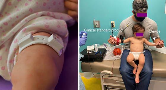
- Successful clinical testing on pediatric patients with cystic fibrosis using a flexible device enabling sweat gland stimulation and simultaneous biosensing. - Two-year collaborative research between KIST and Northwestern University. Sweat contains biomarkers that can monitor various health conditions, from diabetes to genetic disorders. Sweat sampling, unlike blood collection, is preferred by users due to its painless nature. However, to obtain sufficient nutrients or hormones from sweat for testing, intense physical activity was previously required to induce sweat. This method posed challenges for individuals with limited mobility. Dr. Kim Joohee from the Bionics Research Center at the Korea Institute of Science and Technology (KIST, Director Oh Sangrok) and Professor John A. Rogers from Northwestern University jointly announced the development of a convenient sweat monitoring device that does not require physical activity but delivers drug stimulation through the skin. Unlike previous methods that induced sweat through exercise, this device delivers drugs that stimulate sweat glands through the skin. The research team developed a flexible device capable of delivering drugs to sweat glands by applying a current to a hydrogel containing drugs. This device, which is small and soft, can be easily attached to the skin. Sweat induced by the drug is collected in microfluidic channels within the device and analyzed for biomarkers using biosensors. This enables the analysis of biomarkers in sweat, reducing the need for cumbersome hospital visits for testing and lowering the risk of biomarker contamination during testing, thereby increasing accuracy. The device developed by the research team was attached to infants with cystic fibrosis, and the chloride concentration, a biomarker in sweat, was confirmed. The results were consistent with those obtained from traditional analysis methods using sweat collected in hospitals, with an accuracy of over 98%. Additionally, the stability of the device on the skin was ensured by confirming skin temperature and pH values. Since cystic fibrosis mainly manifests during infancy, continuous monitoring of disease progression and physical condition is necessary. With this device, monitoring can be easily done at home, reducing the psychological and physical stress on pediatric patients and their caregivers. This newly developed device contributes to the expansion of non-invasive disease monitoring technology based on sweat in healthy adults as well. Furthermore, the technology of delivering drugs through the skin can be utilized not only to induce sweat but also to increase the delivery rate of drugs in localized areas such as skin conditions or wounds, thereby accelerating recovery. Dr. Kim Joohee stated, "Through two years of collaborative research with Northwestern University, we have not only addressed the limitations of existing methods for inducing sweat but also achieved success in clinical research, bringing us one step closer to commercialization." Professor John A. Rogers added, "We plan to conduct large-scale clinical studies and commercialization, including adults, in the future." [Figure 1] Schematic and Actual Photo of Wearable Device Enabling Drug Delivery for Sweat Induction and Disease Monitoring Illustration and photograph of the device capable of drug delivery for sweat induction and simultaneous monitoring of biomarkers in sweat. [Figure 2] Testing the Wearable Device Attached to a Child A child with the traditional wired device attached to the left arm and the developed device adhered to the right arm, delivering drugs to stimulate sweat glands. [Figure 3] Comparison Graphs of Results and Pain Perception During Testing (Left) Graph showing over 98% agreement between the traditional diagnostic method and the developed device's biomarker analysis results for five patients. (Right) Graph comparing the pain perception experienced by patients during disease monitoring using the traditional diagnostic method and the developed device. The graph indicates that the developed device causes less discomfort compared to the traditional diagnostic method. ### KIST was established in 1966 as the first government-funded research institute in Korea. KIST now strives to solve national and social challenges and secure growth engines through leading and innovative research. For more information, please visit KIST’s website at https://eng.kist.re.kr/ This research was conducted through KIST's major projects and the Outstanding Young Researcher Program (RS-2023-00211342) supported by the Ministry of Science and ICT (Minister Lee Jong-ho). The research findings were recently published online in the latest issue of the international journal "Biosensors & Bioelectronics" (IF 12.6).
- 8
- WriterDr. Kim, Joohee
- 작성일2024.05.28
- Views1601
-
7
Tricking the Brain’s inner GPS: Grid cells responses to the illusion of self-location
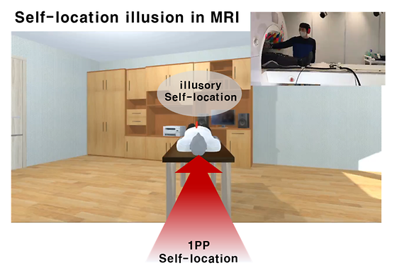
- Researchers have observed grid cell activity in the human brain during self-location illusions induced by multisensory virtual reality, without altering visual perspectives. This groundbreaking study opens up new avenues for the objective diagnosis and treatment of psychiatric symptoms, such as out-of-body experiences, enhancing our understanding of brain mechanisms behind perceptual illusions. Dr. Hyuk-June Moon from the Bionics Research Center at the Korea Institute of Science and Technology (KIST), in collaboration with Prof. Olaf Blanke’s team at the Swiss Federal Institute of Technology Lausanne (EPFL), has successfully induced self-location illusions with multi-sensory virtual reality (VR) in the MRI scanner and observed corresponding changes in the human brain's grid cell activity. The brain is known to contain grid cells and place cells, which perform global positioning system (GPS) functions that allow us to recognize where we are. While traveling to a specific place, the GPS cells along the way fire in turn, depending on their location, and these cells play an important role in recognizing our location in the form of coordinates and remembering events in space. Humans can sometimes perceive themselves to be in a different location without actually moving their physical bodies such as during an illusion, such as out-of-body experience. However, such purely cognitive self-location changes—and the corresponding response of the brain's GPS cells—have not been investigated in animal models like rats, where these perceptions cannot be induced or confirmed. Furthermore, conventional methods to study GPS cell studiess have required opening the skull and measuring the activity of individual cells in the deep brain structures with invasive electrodes, limiting our understanding of human GPS cells. To observe grid cell activity during the illusory self-location changes, the researchers combined MRI-compatible VR technology with multisensory bodily stimulation to induce the illusion, which was precisely controlled in various directions as designed. The fMRI Images measured during the experiment were used to estimate the activities of grid cells, and the subjective illusory experiences of participants were assessed through post-experiment questionnaires and behavioral metrics reflecting their perceived self-location. As a result, the team demonstrated for the first time that purely cognitive changes in magnetic positionsuch illusory self-location changes induced by multisensory bodily stimulation, without any changes in the visual environmental cues, elicit corresponding activities of human grid cells. This is the first clinical study to demonstrate that multisensory bodily stimuli alone can evoke grid cell activities, without any kind of navigation (not active nor imagined) and without change in the visual perspective. It shows that GPS coordinates in the human brain respond not only to the physical location of the body but also to location information based on various cognitive activities and experiences, raising the possibility of objective diagnosis of hallucinatory symptoms through brain image analysis. The findings are also expected to contribute to the development of new therapies by providing targets for the treatment of patients suffering from illusory symptoms such as out-of-body experience. Dr. Moon stated, "Unlike previous human grid cell studies, which have relied on changes in visual environmental cues from a first-person perspective, we have newly suggested a key research element of integrating multisensory bodily signals." adding, "We plan to conduct follow-up international collaborative research to further understand the brain mechanisms underlying illusions caused by various mental and neurological diseases, and to develop non-invasive brain stimulation treatments that can alleviate these symptoms." [Figure 1] Controlled induction of self-location illusion through multisensory VR in the MRI scanner. Combining an MRI-compatible VR system with multisensory (visuo-tactile) bodily stimulation during fMRI scans, illusions that changes perceived self-location illusion were induced in a precisely controlled manner. [Figure 2] Grid cell activities in the Entorhinal cortex during different task conditions Grid cell activities during different task conditions was estimated through fMRI signals in the entorhinal cortex, where grid cells are mostly distributed. Grid cell activity was significantly observed during the illusion condition, where multisensory bodily signals were sychronously integrated, but not in the control condition, where the equivalent level of multisensory signals were not integrated and separately applied. Confirming validity of the methods used in the study, grid cell activity was also observed during normal VR navigation condition. [Figure 3] Similarity between illusion-induced and VR navigation-induced grid cell activity Illusion-induced grid cell activity is significantly correlated with grid cell activity observed during conventional VR navigation with the matched self-location changes (in both direction and distance). This proportional relationship was not observed when illusion induction was not successful (when illusory self-location changes were smaller; < 0.5 meter). This suggests that illusion-induced self-location changes and VR navigation-induced self-location changes evoke similar activity of grid cells. ### KIST was established in 1966 as the first government-funded research institute in Korea. KIST now strives to solve national and social challenges and secure growth engines through leading and innovative research. For more information, please visit KIST’s website at https://eng.kist.re.kr/ This research was supported by the Ministry of Science and ICT (Minister Lee Jong-ho) under the KIST Major Project and the Swiss National Science Foundation (320030_188798). The results of the research were published in March in the international journal PNAS (IF: 11.1).
- 6
- WriterDr. Moon, Hyuk-June
- 작성일2024.05.20
- Views1279
-
5
Developing artificial skin that can regenerate skin and transmit sensation at the same time
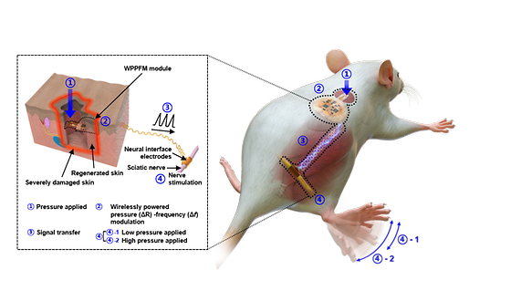
- Development of biomimetic bionic skin and tactile neurotransmission system - Successful animal model implantation of bionic artificial skin composed of sensors and biomaterials Damage to nerve tissue due to skin defects such as burns, skin diseases, and trauma causes loss of sensory and cognitive functions that are essential for life-sustaining activities, as well as mental and physical distress. If the damage is severe enough that natural healing is not possible, surgical treatment is required to implant artificial skin in the affected area, but the artificial skin developed to date has focused on skin regeneration, providing a structure and environment similar to skin tissue, but has not restored sensation to patients. The Korea Institute of Science and Technology (KIST) announced that a research team led by Dr. Youngmee Jung of the Center for Biomaterials and Dr. Hyunjung Yi of the Post-Silicon Semiconductor Institute, in collaboration with Prof. Ki Jun Yu of Yonsei University and Prof. Tae-il Kim of Sungkyunkwan University, has developed a human-implantable tactile smart bionic artificial skin. Unlike conventional artificial skin, which focuses on skin regeneration, smart bionic artificial skin can restore even permanently damaged tactile senses by fusing biocompatible materials and a tactile function delivery system implemented with electronic devices. The artificial skin developed by the team is a hydrogel composed of collagen and fibrin, the main components of skin, that can detect even small pressure changes by inserting crack-based tactile sensors. The sensed pressure changes are converted into electrical signals via Wireless powered pressure-frequency modulation (WPPFM) circuit, which are then transmitted to the nerves by tactile nerve interfacing electrodes, allowing the device to perform the same tactile functions as the skin. The researchers also found that collagen and fibrin, which are responsible for skin's elasticity and tissue connectivity, trigger the proliferation and differentiation of skin cells around the wound to promote skin regeneration. The smart bionic artificial skin was implanted into rats with severe skin damage to test its effectiveness in promoting skin regeneration and reestablishing tactile function, and it showed a wound healing effect of more than 120% compared to the control group at 14 days after implantation. In addition, it detected external changes in the pressure range of 10 to 40 kPa, which is similar to the pressure range felt by human fingertips, and adjusted the electrical signals accordingly to change the rat's response. In particular, the artificial skin developed by the researchers is effective for sensory transmission and skin regeneration because it is implanted directly into the nerves along the subcutaneous fat layer of the damaged skin. After skin regeneration in patients with nerve damage, tactile sensors can operate in the subcutaneous layer, greatly improving independence in daily life. Even in the case of elderly people with degenerated sensory functions, it is expected that the degenerated sensory functions can be restored by directly inserting tactile electronic devices made with high-density integration technology into the subcutaneous layer. "This research is the result of convergence research on devices, materials, and regenerative medicine that effectively combines biomaterials and electronic device technology," said Dr. Youngmee Jung of KIST. "We plan to conduct additional clinical trials in collaboration with medical institutions and companies for commercialization, and we also plan to expand our research to reconstruct various functions of skin tissue such as temperature, vibration, and pain." [Fig 1] Human Implantable Tactile Function Smart Bionic Artificial Skin Components [Fig 2] Neural transmission mechanisms of external stimuli through integrated devices ### KIST was established in 1966 as the first government-funded research institute in Korea. KIST now strives to solve national and social challenges and secure growth engines through leading and innovative research. For more information, please visit KIST’s website at https://eng.kist.re.kr/ This research was supported by the Ministry of Science and ICT (Minister Lee Jong-ho) through the Nano-Materials Source Technology Development Project (2018M3A7B4071106). The findings were published in the latest issue of Nature Communications (IF 16.6, top 7.5% in JCR), a sister journal of Nature and a global authority on international interdisciplinary research.
- 4
- WriterDr. Jung Youngmee
- 작성일2024.04.04
- Views952
-
3
Developing a stem cell therapy to prevent amputations from critical limb ischemia
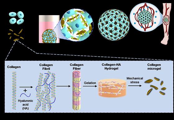
Critical limb ischemia is a condition in which the main blood vessels supplying blood to the legs are blocked, causing blood flow to gradually decrease as atherosclerosis progresses in the peripheral arteries. It is a severe form of peripheral artery disease that causes progressive closure of arteries in the lower extremity, leading to the necrosis of the leg tissue and eventual amputation. Current treatments include angioplasty procedures such as stent implantation and anti-thrombotic drugs, but there is a risk of blood vessel damage and recurrence of blood clots, which is why there is a strong interest in developing a treatment using stem cells. A research team led by Dr. Sangheon Kim of the Center for Biomaterials Research at the Korea Institute of Science and Technology (KIST) announced that they have developed a three-dimensional stem cell therapy to treat critical limb ischemia through a self-assembling platform technology using a new material microgel. By using collagen microgels, a new biocompatible material, the researchers were able to easily transplant stem cells into the body and increase cell survival rate compared to 3D stem cell therapies made of cells alone. Stem cell therapies have high tissue regeneration capabilities, but when stem cells are transplanted alone, hypoxia at the site of injury, immune responses, and other factors can reduce cell viability and prevent the desired therapeutic effect. Therefore, it is necessary to develop a material that delivers stem cells using biodegradable polymers or components of extracellular matrix as a support to increase cell viability. The team processed collagen hydrogels to micro-scale to create porous, three-dimensional scaffolds that are easy to inject in the body and have a uniform cell distribution. Collagen, a component of the extracellular matrix, has excellent biocompatibility and cellular activity, which can induce cell self-assembly by promoting interactions between the microgel particles and collagen receptors on stem cells. In addition, the spacing between microgel particles increased the porosity of the three-dimensional constructs, improving delivery efficiency and cell survival. The microgel-cell constructs developed by the researchers expressed more pro-angiogenic factors and exhibited higher angiogenic potential than cell-only constructs. When microgel-cell constructs were injected into the muscle tissue of mice with critical limb ischemia, blood perfusion rate increased by about 40% and limb salvage ratio increased by 60% compared to the cell-only constructs, confirming their effectiveness in increasing blood flow and preventing necrosis in the ischemic limb. The new stem cell therapy is expected to provide a new alternative for patients with critical limb ischemia who have limited treatment options other than amputation due to its excellent angiogenic effect. Furthermore, since angiogenesis is an essential component of various tissue regeneration processes, it can be extended to other diseases with similar mechanisms to peripheral arterial disease. "The collagen microgel developed in this study is a new biomaterial with excellent biocompatibility and high potential for clinical applications," said Dr. Sangheon Kim of KIST. "We plan to develop technologies for administration methods required in the medical field, as well as conduct follow-up research to clarify the clear mechanism of action of the treatment and discover target factors." [Figure1] Collagen Microgels - The Concept of Self-Assembling Stem Cell Therapy [Figure2] Enhanced cell viability of microgel-assembled stem cell therapeutics [Figure3] Efficacy validation of microgel-cell constructs ### KIST was established in 1966 as the first government-funded research institute in Korea. KIST now strives to solve national and social challenges and secure growth engines through leading and innovative research. For more information, please visit KIST’s website at https://eng.kist.re.kr/ This research was supported by Korean Fund for Regenerative Medicine (22C0620L1). The findings were published in the latest issue of the international journal Bioactive Materials (IF 18.9, JCR top 1.1%). Journal : Bioactive Materials Title : A micro-fragmented collagen gel as a cell-assembling platform for critical limb ischemia repair Publication Date : 2023.12.16. DOI : https://doi.org/10.1016/j.bioactmat.2023.12.008
- 2
- WriterDr. Sangheon Kim
- 작성일2024.03.13
- Views1356
-
1
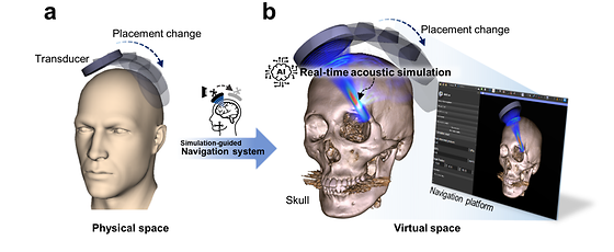
Artificial intelligence lowers the barrier to ultrasound brain disease treatment
- Developing real-time focused ultrasound simulation technology using generative AI models - Expected to improve the accuracy and safety of brain disease treatment with focused ultrasound Focused ultrasound technology is a non-invasive treatment method that focuses ultrasound energy on a few millimeters of the brain, including deep regions, to treat neurological disorders without opening the skull. It has been applied to the treatment of various intractable brain diseases such as depression and Alzheimer's disease because it minimizes the impact on the surrounding healthy tissue and reduces side effects such as complications and infections. However, its use has been limited so far because it is difficult to reflect the distortion of ultrasound waves caused by the different shapes of the skulls of different patients in real-time. A research team led by Dr. Kim, Hyungmin of the Bionics Research Center at the Korea Institute of Science and Technology (KIST) has developed a real-time acoustic simulation technology based on generative AI to predict and correct the distortion of the ultrasound focus position caused by the skull in real-time during focused ultrasound therapy. Until now, the clinical applicability of AI simulation models in the field of non-invasive focused ultrasound therapy technology has not been validated. To predict the location of the invisible acoustic focus, navigation systems based on medical images taken before treatment are currently utilized, which provide information about the relative position of the patient and the ultrasound transducer. However, they are limited by their inability to account for the distortion of ultrasound waves caused by the skull, and while various simulation techniques have been used to compensate for this, they still require significant computational time, making them difficult to apply in actual clinical practice. The research team developed a real-time focused ultrasound simulation technology through an artificial intelligence model based on a generative adversarial neural network (GAN), a deep learning model widely used for image generation in the medical field. The technology reduces the update time of three-dimensional simulation information reflecting changes in ultrasound acoustic waves from 14 s to 0.1 s, while showing an average maximum acoustic pressure error of less than 7% and a focal position error of less than 6mm, both of which are within the error range of existing simulation technologies, increasing the possibility of clinical application. The research team also developed a medical image-based navigation system to verify the performance of the developed technology in order to rapidly deploy it to real-world clinical practice. The system can provide real-time acoustic simulations at the rate of 5 Hz depending on the position of the ultrasound transducer, and succeeded in predicting the position of the ultrasound energy and focus within the skull in real-time during focused ultrasound therapy. Previously, due to the long calculation time, the ultrasound transducer had to be precisely positioned in a pre-planned location to utilize the simulation results. However, with the newly developed simulation-guided navigation system, it is now possible to adjust the ultrasound focus based on the acoustic simulation results obtained in real-time. In the future, it is expected to improve the accuracy of focused ultrasound and provide safe treatment for patients by being able to quickly respond to unexpected situations that may occur during the treatment process. "As the accuracy and safety of focused ultrasound brain disease treatment has been improved through this research, more clinical applications will emerge," said Dr. Kim, Hyungmin of KIST. "For practical use, we plan to verify the system by diversifying the ultrasound sonication environment, such as multi-array ultrasound transducers." [Figure 1] Simulation-Guided Navigation Systems [Figure 2] Clinical Application Examples for Simulation-Guided Navigation ### KIST was established in 1966 as the first government-funded research institute in Korea. KIST now strives to solve national and social challenges and secure growth engines through leading and innovative research. For more information, please visit KIST’s website at https://eng.kist.re.kr/ This research was supported by the Ministry of Science and ICT (Minister Lee Jong-ho) under the Creative Convergence Research Project (CAP-18014-000) of the National Research Council of Korea. The research results were published on October 14 in the top international journal NeuroImage (top 3.6% in JCR). Journal : NeuroImage Title : Real-Time Acoustic Simulation Framework for tFUS: A Feasibility Study Using Navigation System Publication Date : 2023.10.14. DOI : https://doi.org/10.1016/j.neuroimage.2023.120411
- 0
- WriterDr. Kim, Hyungmin
- 작성일2024.01.09
- Views1107
-
-1
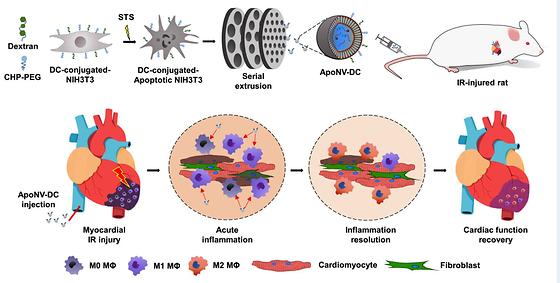
Myocardial infarction, the number one cause of sudden death, may be treated by modulating the immune response.
- Reduced inflammation at the site of myocardial infarction and improved heart function demonstrated - Novel therapy to modulate immune response with apoptotic cell-derived nanovesicles Myocardial infarction, the number one cause of sudden death in adults and the number two cause of death in Korea, is a deadly disease with an initial mortality rate of 30%, and about 5-10% of patients die even if they are transported to a medical center for treatment. The number of myocardial infarction patients in Korea has been increasing steeply, from 99,647 in 2017 to 126,342 in 2021, an increase of 26.8% in five years. Until now, drug administration, percutaneous angioplasty, and arterial bypass surgery have been known as treatments, but they are difficult to apply to severe cases that do not respond to them. Dr. Yoon Ki Joung and Dr. Juro Lee of the Biomaterials Research Center at the Korea Institute of Science and Technology (KIST), together with Prof. Hun-Jun Park and Dr. Bong-Woo Park of the Catholic University of Korea College of Medicine, have developed a new treatment for myocardial infarction that uses nanovesicles derived from fibroblasts with induced apoptosis to modulate the immune response. [Figure 1] SCHEMATIC ILLUSTRATION OF TREATMENT OF MYOCARDIAL ISCHEMIA-REPERFUSION (IR) INJURY WITH THE TARGETED DELIVERY OF APONV-DCS Myocardial infarction is an ischemic heart disease in which the coronary arteries, the blood vessels that supply blood to the heart, become narrowed or blocked, resulting in insufficient blood supply to the heart muscle, which causes nutrient and oxygen deficiency in the myocardium, leading to poor heart function. According to market research firm Technavio, the global myocardial infarction therapeutics market is expected to reach $2.02 billion by 2026, at a CAGR of 4.7%. In recent years, stem cell-derived nanovesicles, such as exosomes, have been used to treat myocardial infarction by modulating the inflammatory response, but stem cells are difficult to produce in large quantities, limiting their economic viability. [Figure 2] Effect on viable myocardium and fibrosis area at 4 weeks after treatment. The research team identified the possibility of treating severe myocardial infarction by reducing the inflammatory response in the heart muscle through a nanomedicine based on apoptotic cells, which are cells that commit suicide due to biochemical changes in their cells. This response was achieved by attaching peptides specific to the site of ischemic myocardial infarction and substances specific to macrophage phagocytosis to the surface of fibroblasts. To this end, the team developed anti-inflammatory nanovesicles that can be delivered specifically to macrophages at the site of myocardial infarction. [Figure 3] Prevention of cardiac function deterioration 4 weeks after the ApoNV-DCs injection. In animal studies, we found that intravenously injected nanovesicles were effectively delivered to the myocardial infarction site in rats and were specifically recruited to macrophages. As a result, the left ventricular ejection fraction, an indicator of the contractile force of the left ventricle, increased by more than 1.5 times compared to the control group for 4 weeks. In addition, the effects of reducing inflammation and fibrosis, and increasing blood vessels preservation rate enhanced cardiomyocytes survival, which resulted in cardiac function improvement. "This is the first study to use nanovesicles produced from apoptosis-induced cells to treat myocardial infarction, and it has the advantage of being able to mass-produce them because it uses other cells rather than stem cells," said Dr. Yoon Ki Joung of KIST. "In the future, we plan to conduct a research to verify the effectiveness and safety of the treatment, including clinical trials, through a collaborative research with Catholic University of Korea Medical School and bio companies." ### KIST was established in 1966 as the first government-funded research institute in Korea. KIST now strives to solve national and social challenges and secure growth engines through leading and innovative research. For more information, please visit KIST’s website at https://eng.kist.re.kr/ This research was supported by the Ministry of Science and ICT (Minister Lee Jong-ho) through the Korea Research Foundation Nano and Material Technology Development Project and the Sejong Science Fellowship Program, and the results were published in the June issue of Advanced Functional Materials (IF:19.0, JCR top 4.7%), an international journal in the field of materials. Journal : Advanced Functional Materials Title : Targeted Delivery of Apoptotic Cell-Derived Nanovesicles prevents Cardiac Remodeling and Attenuates Cardiac Function Exacerbation Publication Date : 02-June-2023 DOI : https://doi.org/10.1002/adfm.202210864
- -2
- WriterDr.Joung, Yoon Ki
- 작성일2023.08.25
- Views1082
-
-3
Safe Bioink for Artificial Organ Printing
- Development of 3D bioprinting ink that induces tissue regeneration without photocuring - Expected applications including patient-specific regenerative treatment technologies, such as artificial organs The development of biomaterials for artificial organs and tissues is active due to an increase in accidental injuries and chronic diseases, along with the entry into a super-aged society. 3D bioprinting technology, which uses cells and biomaterials to create three-dimensional artificial tissue structures, has recently gained popularity. However, commonly used hydrogel-based bioinks can cause cytotoxicity due to the chemical crosslinking agent and ultraviolet light that connect the molecular structure of photocuring 3D-printed bioink. Dr. Song Soo-chang's research team at the Center for Biomaterials, Korea Institute of Science and Technology (KIST, President Yoon Seok-jin), revealed the first development of poly(organophosphazene) hydrogel-based temperature-sensitive bioink that stably maintained its physical structure only by temperature control without photocuring, induced tissue regeneration, and then biodegraded in the body after a certain period of time. [Figure 1] TUNING MECHANICAL PROPERTIES OF BIOINK ACCORDING TO TEMPERATURE AND 3D SCAFFOLD PRINTING Current hydrogel-based bioinks must go through a photocuring process to enhance the mechanical properties of the 3D scaffold after printing, with a high risk of adverse effects in the human body. In addition, there have been possibility of side effects by transplanting externally cultured cells within bioink to increase the tissue regeneration effect. Accordingly, the research team developed a new bioink material using a temperature-sensitive poly(organophosphazene) hydrogel, which existed in a liquid form at low temperatures and changed to a hard gel at body temperature. This enabled the regeneration of tissues only by temperature control without chemical crosslinking agents or UV irradiation and the manufacture of a three-dimensional scaffold with a physically stable structure, minimizing the possibility of immune adverse effects in the human body. [Figure 2] Biodegradation and bone regeneration effects after implanting the 3D-printed scaffold with bioink to the bone-damaged area The developed bioink also had a molecular structure that could interact with growth factors, which were proteins that help in tissue regeneration to preserve growth factors that regulated cell growth, differentiation, and immune responses for a long period of time. The research team was able to maximize the effect of tissue regeneration by creating an environment in which cell differentiation could be autonomously regulated within the 3D scaffold printed with bioink. The research team fabricated the 3D scaffold by printing it with a 3D bioprinter using bioink containing transforming growth factor beta 1 (TGF-β1) and bone morphogenetic protein-2 (BMP-2), which were required for cell infiltration and bone regeneration, and conducted an experiment by implanting it into a damaged bone in a rat. As a result, cells from the surrounding tissue were migrated into the scaffold, and the defected bone was regenerated to a normal tissue level, and the implanted 3D scaffold slowly biodegraded in the body over 42 days. [Figure 3] Image selected as the inside back cover Dr. Song Soo-Chang of KIST said, "The research team has transferred technology for the thermo-sensitive polyphosphazene hydrogel to NexGel Biotech Co., Ltd. in June 2022, and the development of products such as bone graft materials and cosmetic fillers is underway." "As the bioink developed this time has different physical properties, follow-up research to apply it to the regeneration of other tissues besides bone tissue is being conducted, and we expect to finally be able to commercialize bioink tailored to each tissue and organ," he said. ### KIST was established in 1966 as the first government-funded research institute in Korea. KIST now strives to solve national and social challenges and secure growth engines through leading and innovative research. For more information, please visit KIST’s website at https://eng.kist.re.kr/ This research was conducted through the KIST Major Projects supported by the Ministry of Science and ICT (Minister Lee Jong-ho), and the research results were published as the inside back cover in the latest issue of "Small" (IF: 15.153, top 7.101% in the JCR field), an international academic journal in the field of materials. Journal : Small Title : Thermo-Responsive Nanocomposite Bioink with Growth-Factor Holding and its Application to Bone Regeneration Publication Date : 1-Mar-2023 DOI : https://doi.org/10.1002/smll.202203464 Public
- -4
- WriterDr. Song, Soo-chang
- 작성일2023.04.14
- Views2010
-
-5
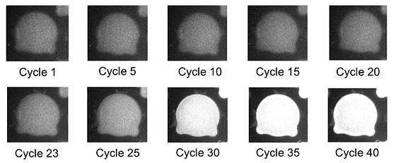
A 5-minute PCR, faster than self-diagnosis kits
- Development of ultrafast PCR technology using materials that generate heat when exposed to light - Expected for rapid diagnosis in pharmacies and clinics due to miniaturized device use PCR technology is a molecular diagnostics technology that detects target nucleic acids by amplifying the DNA amount. It has brought marked progress in the life sciences field since its development in 1984. This technology has recently become familiar to the public due to the COVID-19 pandemic, since PCR can detect nucleic acids that identify the COVID-19 virus. However, due to the technical nature of the PCR test, results cannot be immediately delivered. It takes at least 1 to 2 hours for the test as it requires repeated temperature cycles (60~95℃). Dr. Sang Kyung Kim (Director) and Dr. Seungwon Jung’s research team at the Center for Augmented Safety System with Intelligence, Sensing of the Korea Institute of Science and Technology (KIST, President: Seok Jin Yoon) announced that they had developed an ultrafast PCR technology. By using photothermal nanomaterials, the ultrafast PCR shortens the test time by 10-fold, compared with the time taken for the existing test. The new method is completed in 5 minutes, with diagnostic performance equal to that of the existing test method. Photothermal nanomaterials generate heat immediately upon light irradiation. As such, photothermal nanomaterials rapidly increase in temperature, but it is difficult to maintain performance due to their low stability. The KIST research team has developed a polymer composite that physically holds photothermal nanomaterials and can overcome their instability. By applying it to a PCR system, they have successfully developed a compact PCR system without a heat plate. In addition, they implemented a multiplex diagnostic technology that detects several genes at once, enabling it to distinguish several types of COVID-19 variants in a single reaction. Director Sang Kyung Kim states, “through additional research, we plan to miniaturize the developed ultrafast PCR technology this year, to develop a device that can be utilized anywhere. While maintaining the strength of PCR as an accurate diagnostic method, we will increase its convenience, field applicability, and promptness, by which we expect that it will become a precision diagnostic device that can be used at primary local clinics, pharmacies, and even at home. In addition, PCR technology is a universal molecular diagnostic technology that can be applied to various diseases other than infectious diseases, so it will become more applicable.” This research was carried out by the Practical Convergence Research Center, sponsored by the National Research Council of Science and Technology (Chairperson Bok Chul Kim), and published in the latest online issue of 'ACS Nano' (IF: 18.027, top 5.652% in the JCR field), an authoritative journal in the field of nanomaterials. Journal: ACS Nano Title: Ultrafast Real-Time PCR in Photothermal Microparticles Publication Date: 2022. 12. 6. DOI: https://doi.org/10.1021/acsnano.2c07017 Schematic diagram of PCR temperature cycle using the photothermal effect in polymeric microparticles Changes in fluorescence signals during a real-time PCR of polymeric microparticles
- -6
- WriterDr. Jung, Seungwon
- 작성일2023.02.22
- Views1177
-
-7
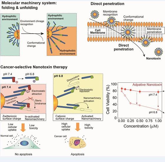
Nanomachines for Direct Penetration of Cancer Cells by Folding and Unfording
- Development of a ‘Nanomachines’ that penetrates and kills cells via mechanical molecular movements - Selective penetration of cancer cells using a latch molecule released near cancer cells Proteins are involved in every biological process, and use the energy in the body to alter their structure via mechanical movements. They are considered biological 'nanomachines' because the smallest structural change in a protein has a significant effect on biological processes. The development of nanomachines that mimic proteins has received much attention to implement movement in the cellular environment. However, there are various mechanisms by which cells attempt to protect themselves from the action of these nanomachines. This limits the realization of any relevant mechanical movement of nanomachines that could be applied for medical purposes. The research team led by Dr. Youngdo Jeong from the Center for Advanced Biomolecular Recognition at the Korea Institute of Science and Technology (KIST, President Seok-Jin Yoon) has reported the development of a novel biochemical nanomachine that penetrates the cell membrane and kills the cell via the molecular movements of folding and unfolding in specific cellular environments, such as cancer cells, as a result of a collaboration with the teams of Prof. Sang Kyu Kwak from the School of Energy and Chemical Engineering and Prof. Ja-Hyoung Ryu from the Department of Chemistry at the Ulsan National Institute of Science and Technology (UNIST, President Yong Hoon Lee), and Dr. Chaekyu Kim of Fusion Biotechnology, Inc. The joint research team focused on the hierarchical structure of proteins, in which the axis of the large structure and the mobile units are hierarchically separated. Therefore, only specific parts can move around the axis. Most existing nanomachines have been designed so that the mobile components and axis of the large structure are present on the same layer. Thus, these components undergo simultaneous movement, which complicates the desired control of a specific part. A hierarchical nanomachine was fabricated by synthesizing and combining 2 nm-diameter gold nanoparticles with molecules that can be folded and unfolded based on the surrounding environment. This nanomachine was comprised of mobile organic molecules and inorganic nanoparticles to function as large axis structures, and defined movement and direction in such a manner that upon reaching the cell membrane, it resulted in a mechanical folding/unfolding movement that led to the nanomachine directly penetrating the cell, destroying the organelles, and inducing apoptosis. This new method directly kills cancer cells via mechanical movements without anticancer medication, in contrast to the capsule-type nanocarriers that deliver therapeutic drugs. Subsequently, a latch molecule was threaded onto the nanomachine to control the mechanical movement to selectively kill cancer cells. The threaded latch molecule was designed to be released only in a low pH environment. Therefore, in normal cells with a relatively high pH (approximately 7.4), the movements of nanomachine was restricted and they could not penetrate the cell. However, at the low pH environment around cancer cells (approximately 6.8), the latch molecules were untied, inducing mechanical movement and cell penetration. Dr. Jeong said, “The developed nanomachine was inspired by proteins that perform biological functions by changing their shape based on their environment. We propose a novel method of directly penetrating cancer cells to kill them via the mechanical movements of molecules attached to nanomachines without drugs. This could be a new alternative to overcome the side effects of existing chemotherapy.” Image Nanomachine, developed by KIST-UNIST joint research team, which selectively penetrates and kills cancer cells, and its mechanism of action The nanomachine directly penetrates cancer cells and kills them by destroying their organelles via mechanical movements of the molecule.
- -8
- WriterDr. Jeong, Youngdo
- 작성일2022.05.16
- Views1340