Brain Science Research Division
-
7
Key to Unlocking the Secret of Degenerative Brain Disorders Found
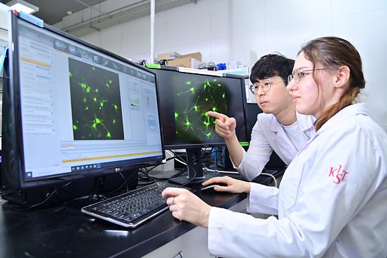
- Development of 'NeuM', a Neuron Labeling Technology Enabling Detailed Observation of Neuronal Structure - Successful Monitoring of Neuronal Changes for up to 72 Hours Alzheimer's disease and Parkinson's disease, along with stroke, are among the top three neurodegenerative disorders, characterized by the malfunction and progressive degeneration of neurons, the nerve cells. Understanding the mechanisms underlying these neurological disorders and developing therapies requires labeling technologies that can visualize neuronal changes not only in normal conditions but also in disease states. A research team led by Dr. Kim Yun Kyung from the Brain Science Institute at the Korea Institute of Science and Technology (KIST), in collaboration with Professor Chang Young-Tae's team from Pohang University of Science and Technology, has announced the development of a next-generation neuron labeling technology called NeuM. NeuM (Neuronal Membrane-selective) selectively labels neuronal membranes, visualizing neuronal structures and allowing real-time monitoring of neuronal changes. Neurons continuously modify their structure and function to transmit information from sensory organs to the brain, regulating thoughts, memories, and behaviors. Therefore, to overcome degenerative neurological diseases, it is essential to develop techniques that selectively label living neurons for real-time monitoring. However, current gene-based and antibody-based labeling technologies, commonly used to observe neurons, suffer from low accuracy and difficulty in long-term tracking due to their dependence on specific gene expression or proteins. NeuM, developed by the research team through molecular design of neuronal cells, possesses excellent binding affinity to neuronal membranes, enabling long-term tracking and high-resolution imaging of neurons. The fluorescent probes within NeuM bind to neuronal membranes utilizing the activity of living cells, emitting fluorescent signals upon excitation by specific wavelengths of light. This visualization of neuronal membranes allows for detailed observation of neuronal terminal structures and high-resolution monitoring of neuronal differentiation and interactions. NeuM, as the first technology to stain cell membranes through endocytosis in living neurons, exhibits selective reactivity towards living cells, excluding dead cells without internalization. Moreover, the research team has succeeded in extending the observation time of neurons from a mere 6 hours to up to 72 hours, enabling the capture of dynamic changes in living neurons over an extended period in response to environmental changes. NeuM is expected to provide insights into research and therapy development for degenerative neurological diseases, for which there are currently no cures. These diseases, including Alzheimer's, result from neuronal damage due to the production of toxic proteins such as amyloid and the influx of inflammatory substances. NeuM's precise observation of neuronal changes can effectively facilitate the evaluation of candidate therapeutic compounds. Dr. Kim stated, "NeuM, developed this time, can distinguish aging and degenerating neurons, becoming a crucial tool in elucidating the mechanisms of degenerative brain disorders and developing treatments." He further added, "In the future, we plan to refine NeuM for even more precise analysis of neurons by designing fluorescence wavelengths to distinguish colors such as green and red." [Figure 1] Molecular Design for Selective Labeling of Neuronal Membranes [Figure 2] Researchers from Dr. Kim Yoon-kyung's team at KIST are utilizing the next-generation neuron labeling technology, 'NeuM,' to visualize neurons in real-time and examine high-resolution images. ### KIST was established in 1966 as the first government-funded research institute in Korea. KIST now strives to solve national and social challenges and secure growth engines through leading and innovative research. For more information, please visit KIST’s website at https://eng.kist.re.kr/ This research was supported by the Ministry of Science and ICT (Minister Lee Jong-ho) through KIST's major projects and the Dementia Overcoming Project (RS-2023-00261784). The research results have been published in the latest issue of the international academic journal "Angewandte Chemie."
- 6
- WriterDr. Kim Yun Kyung
- 작성일2024.04.08
- Views1138
-
5
New treatment developed to dramatically slow down the progression of blindness-causing retinal diseases:
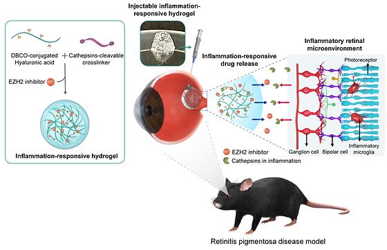
- Interactive release of anti-inflammatory drugs depending on the level of retinal degeneration. - A customized treatment approach is expected to be developed to reduce patients’ inconvenience of having multiple shots. The Korea Institute of Science and Technology (KIST) announced that Dr. Maesoon Im of the Brain Science Institute, together with Prof. Seung Ja Oh of Kyung Hee University and Prof. Kangwon Lee of Seoul National University, successfully incorporated anti-inflammatory drugs into a hydrogel to suppress inflammation in the retina and effectively deliver the drugs to the inflamed area. Age-related macular degeneration and retinitis pigmentosa are incurable eye diseases that cause blindness due to the gradual damage of photoreceptor cells, which convert light into biological signals in the retina, the light-sensitive tissue at the back of the eye. Age-related macular degeneration is a condition that damages the macula, the central part of the retina, and is the number one cause of blindness in people over the age of 65. Retinitis pigmentosa, on the other hand, is a genetic disorder that causes gradual death in the photoreceptor cells in the retina and affects about 1 in 4,000 people worldwide, initially causing night blindness but eventually leading to vision loss. Currently, there is no effective cure for either disease, and one of the treatments is to inject anti-inflammatory drugs into the eye to slow down the degree of retinal damage. However, these injections only work for as long as the drug remains in the eye, requiring patients to visit a clinic for intraocular injections every four to 12 weeks, depending on how long the effect of the drug lasts. For the first time, the team utilized a substance that inhibits the inflammatory factor EZH2, which contributes to retinal degeneration, along with an anti-inflammatory agent. When mice with retinal degeneration were injected with the anti-inflammatory drug, the progression of retinal degeneration slowed down. The researchers have successfully developed a hydrogel that slowly degrades upon encountering the enzyme cathepsin, which is typically overexpressed in inflammatory environments, to deliver anti-inflammatory drugs. When the team's drug-loaded inflammation-responsive hydrogel was injected into the eyes of mice suffering from retinal degeneration, inflammatory factors in the retina were reduced to approximately 6.1%. The team also found that the protective effect on photoreceptor cells, which are known to be destroyed by retinal degeneration, was about four times higher than in the control group, effectively delaying vision loss. Notably, the hyaluronic acid-based hydrogel, which has similar mechanical and optical properties to the vitreous humor of the eye, allows for different rates of hydrogel degradation in each patient, minimizing the need for repeated injections. This newly developed technology is expected to reduce the economic burden and the risk of accidents during outpatient visits for patients with difficulty in mobility due to visual impairment. Additionally, for patients in the early stages of symptoms, reducing the frequency of hospital visits can alleviate inconvenience in daily life. "For future commercialization, we plan to digitize the amount of drug and hydrogel used, as well as the treatment period, according to the progression of the disease. We also intend to assess the long-term stability of the drug delivery system," said Dr. Maesoon Im of KIST. "In addition to the retinal degenerative diseases, we will investigate inflammation levels in other retinal diseases to see if our inflammation-responsive drug delivery system would work on those conditions," said Prof. Seung Ja Oh of Kyung Hee University. [Figure 1] Schematic illustration of syringe-injectable inflammation-responsive hydrogel for suppression of inflammatory microglia for preventing photoreceptor death in retinitis pigmentosa. [Figure 2] The effectiveness of the developed inflammation-responsive drug was verified. ### KIST was established in 1966 as the first government-funded research institute in Korea. KIST now strives to solve national and social challenges and secure growth engines through leading and innovative research. For more information, please visit KIST’s website at https://eng.kist.re.kr/ This research was supported by the Ministry of Science and ICT (Minister Lee Jong-ho) through the KIST Major Project and Young and Mid-Career Researchers, the Excellent Young Researcher Support Project, the Brain Function Identification and Regulation Technology Development Project, and the Public Benefit Medical Technology Research Project of the Ministry of Health and Welfare (Minister Cho Kyu-hong). The research were published in the latest issue of the international journal 'npj Regenerative Medicine' (IF 7.2, top 19.3% in JCR). Journal : npj Regenerative Medicine Title : Effective Protection of Photoreceptors Using an Inflammation-Responsive Hydrogel to Attenuate Outer Retinal Degeneration Publication Date : 2023.12.14. DOI : https://doi.org/10.1038/s41536-023-00342-y
- 4
- WriterDr. Im Maesoon
- 작성일2024.02.14
- Views1584
-
3
Capturing Nanoplastics in Tap Water with Light
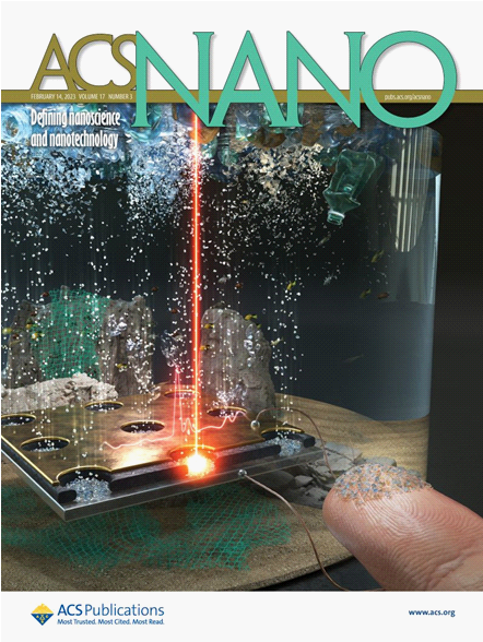
- Electro-photonic tweezer captures and detects trace amount of nanoplastics through surface-enhanced Raman scattering - Application in safe water resource management technology Nanoplastics are plastics that have been discarded from our daily lives and that enter ecosystems in the size scale below 1 micro-metter after their physical and chemical disintegration. Recent research has shown that the concentration of microplastics in the major rivers in South Korea is the highest worldwide; it is not unusual to find news reports about the detection of microplastics in simple tea bags or drinking water and nanoplastic is the worse. The impact of micro/nanoplastics on human health and the environment in general is considered to be significant. However, the detection of nanoplastic is limited because their concentration is low and their size is extremely small. In addition, the detection process requires a few hours to days, and incurs significant costs during the pre-processing step of concentrating the plastic sample. The research team of Dr. Yong-sang Ryu at the Brain Research Institute of the Korea Institute of Science and Technology (KIST) used an electro-photonic tweezer along with metal nanoparticles to concentrate ultrafine nanoplastics within a short period, and they reported the development of a real-time detection system using light. The research team supplied electricity to a large-area vertically-aligned metals sandwiched by nanofilm insulator. They conducted Raman spectroscopy, which analyzes the energy difference between the incident and scattered light according to the frequency of the molecule. By combination of two technique: electrical nanoparticle capture together with real-time Raman spectroscoy, the research team achive in-situ the detection of a 30-nm 10 μg L-1 polystyrene particle with the help of gold nanoplarticles via Surface-enhanced Raman spectroscopy. Moreover, the research team easily separated the particle from the sample through the dielectrophoresis phenomenon. Thus, the entire process including the collection, separation, and analysis for nanoplastics analysis, , which previously required at least one day, was drastically reduced to several seconds by employing an original technology that separates and detects plastics using one platform. Researchers Euitae Jeong and Dr. Eui-Sang Yu (common lead author) at KIST, who performed this research, reported that "the findings of this research are meaningful in that ultrahigh-sensitivity detection of microplastics in real-time has become possible, and the proposed approach can be extended to the measurement of the microplastic concentration in various water resources and applied as a water resource securement technology.“ This research was carried out as a major project of KIST with the support of the Ministry of Science and ICT (Minister Jongho Lee); the results have been reported as a cover paper in the latest international journal 「ACS Nano」 (IF: 18.027). Journal: ACS Nano Title: Real-time Underwater Nanoplastic Detection Beyond Diffusion Limit and Low Raman Scattering Cross-section via Electro-photonic Tweezers Publication Date: 27-Dec-2022 DOI: https://doi.org/10.1021/acsnano.2c07933 ACS Nano front cover selection Raman-spectroscopy-based nanoplastic detection using the electric-optical tweezer and via surface-enhanced Raman scattering and the subsequent amplification of optical signals as well as the reduction of the accumulation time. Top right: Mimetic diagram of subsequent accumulation time reduction (blue: existing, red: current research) Bottom right: Mimetic diagram of subsequent amplification of optical signal accordingly (blue: existing, red: current research)
- 2
- WriterDr. Ryu, Yong-Sang
- 작성일2023.02.27
- Views1364
-
1
Development of Artificial Synaptic Semiconductor Device Based on New 2D Materials
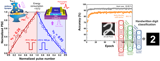
- Utilization of new two-dimensional materials as thin as a single atomic layer - Development of semiconductor devices that operate at low energy like human synapses The Korea Institute of Science and Technology (KIST, President Yoon Seokjin) announced that the research team of Dr. Joon Young Kwak at the Artificial Brain Convergence Research Center developed a new two-dimensional insulator material synthesis technology with a new element composition ratio, as well as a high-performance and low-power artificial synaptic semiconductor device using this new material, through joint research with Professor Ki-beom Kang's team at the Korea Advanced Institute of Science and Technology (KAIST) and Dr. Taek-mo Jeong's team at the Korea Research Institute of Chemical Technology (KRICT). With the recent increase in the proportion of video and image data, the processing of unstructured data is drawing attention as a key factor in the development of future artificial intelligence (AI) systems. In line with this trend, to overcome the excessive power consumption and limited information processing performance of the current widely used von Neumann computing structure, a “neuromorphic system” that can process and learn information with high efficiency and low power consumption is emerging as a next-generation semiconductor system. Neuromorphic systems mimic the human brain to increase computing performance while reducing power consumption. To implement this, it is necessary to develop high-performance next-generation semiconductor devices that can precisely simulate “synapses” that regulate the connection strength between neurons according to the input signals. Silicon-based semiconductor devices, which are predominantly used at present, consume much more energy than biological synapses, and have physical limitations in simulating a highly integrated system similar to a real nervous system. For this reason, research is actively being conducted to realize high-performance artificial synaptic devices by applying the properties of materials such as oxides and organic/inorganic materials. In addition, newly emerging two-dimensional materials are very thin at the atomic level, which gives them a great advantage in high integration of semiconductor devices. They have superior performance compared to existing silicon materials, such as fast switching speed and charge transfer speed, due to their unique characteristics. The joint research team developed a synaptic device based on a new 2D insulator material and a heterojunction structure of a 2D semiconductor, enabling electrons to move efficiently even at low energy. Using these physical characteristics, they succeeded in developing an artificial synaptic device that shows uniform synaptic connection strength change and operates with an energy of about 15 fJ, which is similar to the actual energy consumption of human synapses. In addition, synaptic connection strength can be maintained for a short or long time depending on the number and intensity of external stimuli, enabling more precise simulation of human brain functions. The research team attempted artificial intelligence learning based on the developed high-performance two-dimensional artificial synaptic device, and the classification accuracy of handwritten digit image data (MNIST) was about 88.3%, confirming the possibility of application to actual neuromorphic systems. Dr. Kwak of KIST said, “As the importance of research on high-efficiency new materials that can be used as substitutes for silicon in the development of next-generation semiconductors is growing, synaptic devices based on the heterojunction structure of semiconductors and the new two-dimensional insulator material presented in this study should have excellent competitiveness in implementing high-level neuromorphic hardware that can accurately simulate brain behavior." This research was carried out with the support of a KIST institutional research program, the Next-Generation Intelligent Semiconductor Technology Development Project of the National Research Foundation of Korea, and the New Concept PIM Semiconductor Leading Technology Development Project of the National Institute of Information and Communications Technology Evaluation. The research results were published in the latest issue of the international journal Advanced Materials (IF: 32.086). [Core Figure (Main)] Characteristics of the low-power, high-performance artificial synapse (left) and image classification learning accuracy test (right) of the new 2D-material-based artificial synaptic device developed by the research team. [Reference figure] Synthesis technology developed by the research team and structure and analysis of the new 2D material.
- 0
- WriterDr. Kwak, Joon Young
- 작성일2022.10.10
- Views1315
-
-1
Discovery of the Causes of Brain Dysfunction in Patients with Huntington’s Disease
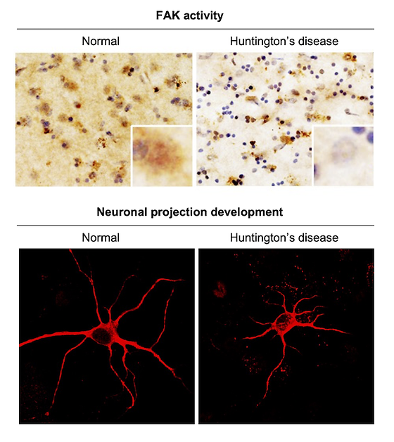
- A protein crucial to synaptic function in brain tissues of patients with Huntington’s disease (HD) was discovered to have decreased function by researchers at KIST Huntington’s disease (HD) is a hereditary brain disease caused by a mutation in the huntingtin gene. HD is a neurodegenerative disease without a cure that, after the onset of the disease at around 40 years of age, causes changes in personality and symptoms of dementia along with uncontrollable convulsive movements, ultimately leading to death. It is known that such HD symptoms are caused by the destruction of brain cells in the striatum due to problems occurring in synapses that are crucial to brain function during the progression of the disease. However, the specific mechanism behind brain dysfunction during the progression of HD has not been fully elucidated. The research team lead by Dr. Jihye Seong and Dr. Hoon Ryu, principal researchers at the Brain Science Institute (BSI) of Korea Institute of Science and Technology (KIST, President Seokjin Yoon), was said to have found significantly reduced activity of focal adhesion kinase (FAK) proteins that play an important role in neurite motility and proper synapse formation in the brain tissues of patients with HD. Activated FAK proteins play an important role in brain function as they are essential in neurite motility and proper synapse formation. The KIST research team identified a significant reduction in FAK activity in HD cells and mouse models, as well as brain tissues of HD patients. These results were also verified through accurate measurements of FAK activity in live cells using a fluorescence resonance energy transfer (FRET)-based biosensor. Phosphatidylinositol 4,5-biphosphate (PIP2), a phospholipid found in the cell membrane, is essential for the activation of FAK proteins. Using super-resolution structured illumination microscopy, the research team found that PIP2 in HD cells was unusually strongly bound to the mutant huntingtin protein, inhibiting proper distribution of PIP2 throughout the cell membrane. This abnormal distribution of PIP2 inhibits FAK activation, which hinders proper synaptic function, causing brain dysfunction in the early stages of HD. Dr. Seong said, “The pathological mechanisms of synaptic dysfunction in patients with Huntington’s disease revealed through this study could be utilized as a therapeutic target for the treatment of brain dysfunction.” Dr. Ryu said, “Because the results of this study show the pathological mechanisms found in actual brain tissues of patients with HD, I believe it has a greater significance in suggesting a new therapeutic target for human degenerative brain diseases.” Image [Figure 1] Differences in FAK activation and neuronal protrusion formation in brain tissues of normal and Huntington's disease patients [Figure 2] Inhibition of FAK activation due to abnormal distribution of phospholipid caused by mutant huntingtin
- -2
- WriterDr. Seong, Jihye
- 작성일2022.09.16
- Views1491
-
-3
Cancer Immunotherapy Capable of Modulating Tumor Immunophenotypes
- Small-molecule activation of innate immunity induces the infiltration of immune cells into cancer cells - Expected applications include various combination therapies for immuno-oncology Innovations in cancer immunotherapy have achieved clinical success by considerably increasing the survival rate of patients undergoing cancer treatment. However, there still exists an unmet medical need due to the low response rate to checkpoint inhibitors caused by the low immune reactivity of cancer cells in “cold” tumors. In their efforts to turn “cold” tumors into “hot” tumors, many global pharmaceutical companies have been focusing on utilizing the innate immune regulatory protein known as STING to increase the immunoreactivity of tumors and the infiltration of immune cells into the tumor microenvironment (TME). However, since clinical trials on the first STING agonist, ADU-S100, were suspended in 2020, there is an urgent need to develop new STING activators. Under these circumstances, a research team led by Dr. Sanghee Lee of the Brain Science Institute at the Korea Institute of Science and Technology (KIST; President: Seok-Jin Yoon), and Dr. Hyejin Kim of the Infectious Diseases Therapeutic Research Center at the Korea Research Institute of Chemical Technology (KRICT; President: Mihye Yi) announced the development of a new small-molecule STING agonist. Once the STING agonist was stimulated by a compound, it induced the secretion of cytokines such as interferons (IFNs) and activated an innate immune response mediated by T cells. The activated immune system altered the immune phenotype of the tumor, turning it from “cold” with a low reactivity to T cells to “hot” with a high reactivity, leading to the recruitment of T cells in the TME. In this study, compound administration effectively inhibited the growth of cancer cells in mice models. In particular, 20% of the treated group was found to be tumor-free as a result of the complete elimination of their tumors. Furthermore, immunological memory suppressed the growth of recurrent tumors without need for additional drug administration. Ultimately, no tumor growth was observed in the tumor-free group after the first treatment. Most of the existing STING agonists were subjected to intratumoral administration, which limited the broad application of cancer treatment, whereas the compound in this study was able to be administered by intravenous injection. In terms of further drug development, this agent is also able to be applied to combination cancer therapies and current standard treatments, such as radiation therapy, chemotherapy, and monotherapy. Dr. Lee stated, “Everyone dreams of vanquishing cancer; however, the development of cancer immunotherapeutics for ailments such as brain tumors is still limited. We hope that this study can provide the seeds for new therapeutic strategies for cancers where immunotherapy has had limited application.” Image Supplementary cover image of J. Med. A schematic diagram in which the substance developed in this study stimulates immune cells, activates an innate immune response, and induces cancer cell death. Chemical structure and mechanism of action of the STING agonist with the 4c compound developed in this study (left) and results and schematic diagram of anticancer efficacy in animal models (right).
- -4
- WriterDr. Lee, Sanghee
- 작성일2022.05.31
- Views1550
-
-5
5 to over 50 Days’ Significant Improvement in 10㎚ Thick Artificial Cell Membrane Stability
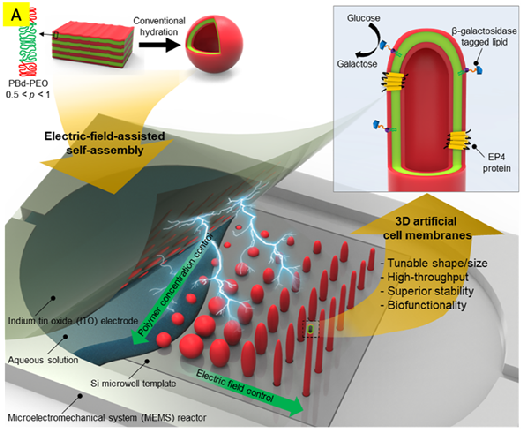
- New achievements of a durable cell mimic thin membrane structure - Tunable and controllable cell-like 3D shapes fabrication on a silicon substrate on-demand: New momentum for future biosensor In nature, the cell membrane has a unique function of protecting the internal from the external environment and communicating outside by sensing the external chemical or physical stimuli like the most precise biosensor for life. The cell membrane, which contains a hydrophilic part that is miscible well with water on the one side and a hydrophobic part that is not miscible well with water on the other, opens and closes ion channels like a water faucet and converts a physicochemical stimulus into an electrical signal which is then transmitted to cells. Active research worldwide on biosensors that can mimic the cell membrane’s excellent sensing has been suggested. However, till recently, the limited ability of an artificial cell membrane structure to only last a maximum of 5 days has been a hurdle. The Korea Institute of Science and Technology (KIST, President Seok-Jin Yoon) announced that the research team led by Dr. Tae Song Kim of the Brain Science Institute has succeeded in developing an artificial cell membrane that can be kept stable for over 50 days on a silicon substrate. This is the longest time reported in the field. In addition to creating, in 2018, an artificial cell membrane lasting for five days, in 2019, Dr. Kim’s team demonstrated the transfer of a positive ion to the inside of a structure with an artificial cell membrane with a protein attached to the surface, confirming its biosensor application potential. However, the durability of at least one month is essential for life science research utilizing artificial cell membranes and the practical commercialization of biosensors. To extend the limit of 5 days of stability of an artificial cell membrane, the KIST research team focused on a material called block copolymer (BCP). A BCP is a macromolecule consisting of two or more blocks, which can be repeatedly aligned as a long row of blocks of counteracting properties that mimic the hydrophilic and hydrophobic nature of the human cell membrane. Dr. Kim’s research team developed a technology that regularly arranges tens of thousands of holes with a diameter of 8 μm (micrometer) on a silicon substrate and inserts a specific amount of BCP solution into each hole through surface treatment, and dries it. Then, a soap bubble-shaped, an elongated oval-shaped, or a thin tubular-shaped BCP double-layer structure is tunably created by applying an electric field between the upper plate electrode of the microfluidic channel and the lower silicon substrate. This process led to the discovery of the possibility of maintaining a structure with a specific shape depending on the concentration of the solution and the applied electric field and frequency. This suggests a means to freely control the size and shape of artificial cell membranes, from a sphere, like a soap bubble, to a cylinder, like a tube. The KIST research team finally created an artificial cell membrane that can be kept stable for over 50 days by filling the outside of a three-dimensional double-layered BCP structure with a porous hydrogel that exhibits excellent elasticity and resilience characteristics similar to that of a human body substance. In addition, an artificial organ structure was produced by replicating an epithelial cell in the small intestine, which consists of thousands of tubular structures (cilia) using a BCP double-layered structure, proving its usage potential as a material for artificial organs through binding with β-galactosidase. Dr. Kim from KIST said, “While global research on artificial cell membranes has been focusing on placing a two-dimensional planar structure on a silicon substrate, the team has succeeded in extending the stability period of an artificial cell membrane by more than ten times following the development of the first three-dimensional artificial cell membrane structure fabrication technology,” and added, “The research, which has presented a path for large area array fabrication of artificial cell membranes, is expected to further develop into a platform technology for biological functionality research that identifies the roles of ultra-sensitive biosensors resembling cell functions, drug screening for new drug development, and neurotransmitters and hormones in the brain.” Image Schematic diagram of manufacturing double-layer structures of various sizes and shapes by controlling the concentration and electric-field of the block copolymer (PBd-PEO) by applying electric fields to the upper and lower layers of the substrate Numerous fabricated spherical and tubular structures and a lateral confocal photomicrograph of a single structure Size distribution of each spherical and tubular structure
- -6
- WriterDr. Kim, Tae Song
- 작성일2022.05.18
- Views1367